Growth and Surface Structure of Fe Monolayers on Pt(110)
Authors
-
Olzat Toktarbaiuly
 Ph.D., National Laboratory Astana, Nazarbayev University, Astana, Kazakhstan
-
Nazerke S. Kydyrbay
Master (Sci.), National Laboratory Astana, Nazarbayev University, Astana, Kazakhstan
-
Mergen R. Zhazitov
National Laboratory Astana, Nazarbayev University, Astana, Kazakhstan
-
Yerbolat Zh. Tezekbay
Master (Sci.), National Laboratory Astana, Nazarbayev University, Astana, Kazakhstan, Kazakhstan
-
Alshyn Zh. Abduvalov
Ph.D., Associate Professor, School of Intelligent Systems, Astana IT University, Astana, Kazakhstan
-
Timur M. Serikov
Ph.D., Associate Professor, Buketov Karaganda National Research University Karaganda, Kazakhstan
DOI:
https://doi.org/10.31489/2026N3/16-27Keywords:
Fe growth, Pt(110), surface reconstruction, epitaxial growthAbstract
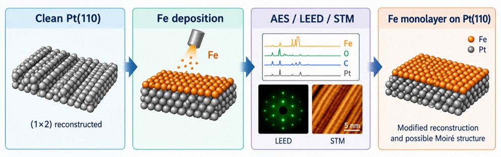
The structural evolution of Fe monolayers deposited on a Pt(110) surface was investigated under ultra-high vacuum conditions using scanning tunneling microscopy, low-energy electron diffraction, and Auger electron spectroscopy. A clean Pt(110) substrate exhibiting the characteristic (1×2) missing-row reconstruction was prepared by repeated cycles of argon ion sputtering, oxidation, and high-temperature annealing. Surface cleanliness and reconstruction were confirmed by Auger electron spectroscopy and low-energy electron diffraction, while scanning tunneling microscopy imaging revealed an anisotropic terrace–step morphology with atomic rows characteristic of the reconstructed Pt(110) surface. Fe monolayers were subsequently deposited on the clean substrate at room temperature by electron-beam evaporation. Auger electron spectroscopy measurements confirmed the presence of iron on the surface, together with minor carbon and oxygen contamination. Low-energy electron diffraction patterns recorded after Fe deposition showed a noticeable modification of the substrate diffraction features, indicating partial suppression of the original Pt(110)-(1×2) reconstruction. In addition, a sixfold diffraction feature observed at higher electron energy suggests the formation of an ordered overlayer or Moiré-type superstructure arising from the lattice mismatch between Fe and Pt(110). These results provide insight into the growth behavior and interface structure of Fe monolayers on reconstructed Pt(110), and establish a basis for future studies of iron oxide formation and redox transformations on this surface.
References
Somorjai, G. A., & Li, Y. (2010). Introduction to surface chemistry and catalysis (2nd ed.). John Wiley & Sons. https://www.wiley-vch.de/en/areas-interest/natural-sciences/introduction-to-surface-chemistry-and-catalysis-978-0-470-50823-7
Freund, H.-J., Kuhlenbeck, H., & Staemmler, V. (1996). Oxide surfaces. Reports on Progress in Physics, 59(3), 283–347. https://doi.org/10.1088/0034-4885/59/3/002 DOI: https://doi.org/10.1088/0034-4885/59/3/001
Campbell, C. T. (1997). Ultrathin metal films and particles on oxide surfaces: Structural, electronic and chemisorptive properties. Surface Science Reports, 27(1–3), 1–111. https://doi.org/10.1016/S0167-5729(96)00011-8 DOI: https://doi.org/10.1016/S0167-5729(96)00011-8
Freund, H.-J., & Pacchioni, G. (2008). Oxide ultra-thin films on metals: New materials for the design of supported metal catalysts. Chemical Society Reviews, 37(10), 2224–2242. https://doi.org/10.1039/B718768H DOI: https://doi.org/10.1039/b718768h
Diebold, U. (2003). The surface science of titanium dioxide. Surface Science Reports, 48(5–8), 53–229. https://doi.org/10.1016/S0167-5729(02)00100-0 DOI: https://doi.org/10.1016/S0167-5729(02)00100-0
Wintterlin, J., & Bocquet, M.-L. (2009). Graphene on metal surfaces. Surface Science, 603(10–12), 1841–1852. https://doi.org/10.1016/j.susc.2008.08.037 DOI: https://doi.org/10.1016/j.susc.2008.08.037
Vlieg, E., Robinson, I. K., & Kern, K. (1990). Relaxations in the missing-row structure of the (1×2) reconstructed surfaces of Au(110) and Pt(110). Surface Science, 233(3), 248–254. https://doi.org/10.1016/0039-6028(90)90636-M DOI: https://doi.org/10.1016/0039-6028(90)90636-M
Sowa, E. C., Van Hove, M. A., & Adams, D. L. (1988). The missing-row model for the reconstructed Pt(110)-(1×2) surface: A LEED intensity analysis showing multilayer distortions. Surface Science, 199(1–2), 174–182. https://doi.org/10.1016/0039-6028(88)90406-2 DOI: https://doi.org/10.1016/0039-6028(88)90406-2
McIntyre, B. J., Salmeron, M., & Somorjai, G. A. (1993). In situ scanning tunneling microscopy study of platinum (110) in a reactor cell at high pressures and temperatures. Journal of Vacuum Science & Technology A, 11(4), 1964–1968. https://doi.org/10.1116/1.578531 DOI: https://doi.org/10.1116/1.578531
Minca, M., Penner, S., Loerting, T., Menzel, A., Bertel, E., Zucca, R., & Redinger, J. (2007). Chemisorption of hydrogen on the missing-row Pt(110)-(1×2) surface. Topics in Catalysis, 46(1–2), 161–167. https://doi.org/10.1007/s11244-007-0326-4 DOI: https://doi.org/10.1007/s11244-007-0326-4
Fery, P., Moritz, W., & Wolf, D. (1988). Structure determination of the (1×2) and (1×3) reconstructions of Pt(110) by low-energy electron diffraction. Physical Review B, 38(11), 7275–7284. https://doi.org/10.1103/PhysRevB.38.7275 DOI: https://doi.org/10.1103/PhysRevB.38.7275
Doná, E., Cordin, M., Deisl, C., Bertel, E., Franchini, C., Zucca, R., & Redinger, J. (2009). Halogen-induced corrosion of platinum. Journal of the American Chemical Society, 131(8), 2827–2829. https://doi.org/10.1021/ja809674n DOI: https://doi.org/10.1021/ja809674n
Pedersen, T. M., Li, W.-X., & Hammer, B. (2006). Structure and activity of oxidized Pt(110) and α-PtO₂. Physical Chemistry Chemical Physics, 8(13), 1566–1574. https://doi.org/10.1039/B515166J DOI: https://doi.org/10.1039/b515166j
Pendry, J. B. (1974). Low-energy electron diffraction. In Interaction of atoms and molecules with solid surfaces (pp. 201–211). Springer. https://doi.org/10.1007/978-1-4684-8777-0_7 DOI: https://doi.org/10.1007/978-1-4684-8777-0_7
Briggs, D., & Seah, M. P. (Eds.). (1990). Practical surface analysis, Volume 1: Auger and X-ray photoelectron spectroscopy (2nd ed.). John Wiley & Sons. ISBN 0-471-92081-9.
Chen, C. J. (2021). Introduction to scanning tunneling microscopy. Oxford University Press. https://academic.oup.com/book/41125 DOI: https://doi.org/10.1093/oso/9780198856559.001.0001
Bode, M. (2003). Spin-polarized scanning tunnelling microscopy. Reports on Progress in Physics, 66(4), 523–582. https://doi.org/10.1088/0034-4885/66/4/203 DOI: https://doi.org/10.1088/0034-4885/66/4/203
Spiridis, N., Wilgocka-Ślęzak, D., Freindl, K., Figarska, B., Giela, T., Młyńczak, E., & Korecki, J. (2012). Growth and electronic and magnetic structure of iron oxide films on Pt(111). Physical Review B, 85(7), Article 075436. https://doi.org/10.1103/PhysRevB.85.075436 DOI: https://doi.org/10.1103/PhysRevB.85.075436
Ritter, M., Ranke, W., & Weiss, W. (1998). Growth and structure of ultrathin FeO films on Pt(111) studied by STM and LEED. Physical Review B, 57(11), 7240–7247. https://doi.org/10.1103/PhysRevB.57.7240 DOI: https://doi.org/10.1103/PhysRevB.57.7240
Lewandowski, M., Pabisiak, T., Michalak, N., Miłosz, Z., Babačić, V., Wang, Y., Korecki, J., & Kiejna, A. (2018). On the structure of ultrathin FeO films on Ag(111). Nanomaterials, 8(10), Article 828. https://doi.org/10.3390/nano8100828 DOI: https://doi.org/10.3390/nano8100828
Shipilin, M., Lundgren, E., Gustafson, J., Zhang, C., Bertram, F., Nicklin, C., & Merte, L. R. (2017). Fe oxides on Ag surfaces: Structure and reactivity. Topics in Catalysis, 60(6–8), 492–502. https://doi.org/10.1007/s11244-016-0714-8 DOI: https://doi.org/10.1007/s11244-016-0714-8
Soldemo, M., Niu, Y., Zakharov, A., Lundgren, E., & Weissenrieder, J. (2015). A well-ordered surface oxide on Fe(110). Surface Science, 639, 13–19. https://doi.org/10.1016/j.susc.2015.04.008 DOI: https://doi.org/10.1016/j.susc.2015.04.008
Seidel, P., Pomp, S., Schwarz, F., & Sterrer, M. (2025). Stability and dissolution of single-crystalline iron oxide thin films in electrochemical environments. Surface Science, 751, Article 122621. https://doi.org/10.1016/j.susc.2024.122621 DOI: https://doi.org/10.1016/j.susc.2024.122621
Venables, J. A. (1994). Atomic processes in crystal growth. Surface Science, 299–300, 798–817. https://doi.org/10.1016/0039-6028(94)90698-X DOI: https://doi.org/10.1016/0039-6028(94)90698-X
Brune, H. (1998). Microscopic view of epitaxial metal growth: Nucleation and aggregation. Surface Science Reports, 31(4–6), 125–229. https://doi.org/10.1016/S0167-5729(99)80001-6 DOI: https://doi.org/10.1016/S0167-5729(99)80001-6
Downloads
Published online
How to Cite
Issue
Section
License
Copyright (c) 2026 Eurasian Physical Technical Journal

This work is licensed under a Creative Commons Attribution-NonCommercial-NoDerivatives 4.0 International License.